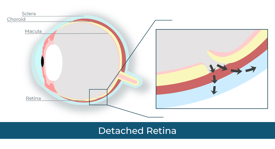
Symptoms of retinal detachment include:
- A gray curtain covering some of your field of vision
- A shadow in your side vision
- Seeing flashing lights
- Seeing many new floaters
What are the Treatment Options for a Detached Retina?
There are five different surgical options for a detached retina. The type of treatment your doctor suggests will depend on how severe the damage is and what type of retinal detachment you have.
Freeze Treatment (Cryopexy)
Retinal cryopexy is a less invasive treatment option that a doctor may suggest if the tear in your retina is very small. It can often be done in the doctor’s office.
Your doctor will first put numbing medicine in your eye. Then they will take a small freezing probe and touch the white outside part of your eye, called the sclera, in the area right over the tear. The cold from the probe causes scar tissue to form around the tear, which holds the retina in place.
Laser Surgery (Photocoagulation)
Photocoagulation is a type of retina surgery that uses a medical laser. Like freeze treatment, your doctor may suggest it for small tears, and it can usually be done in a doctor’s office.
In laser surgery, your doctor will numb your eye, then shine a medical laser inside your eye. The laser makes small burns in your retina that create scar tissue around the hole. This seals the hole and holds the retina in place.
Pneumatic Retinopexy
In a pneumatic retinopexy, your doctor will use an air bubble to push your retina back into place. They then use freeze treatment or laser surgery to repair any tears in the retina.
To perform a pneumatic retinopexy, your doctor will first numb your eye. They’ll then insert a very small needle into your eye and remove a bit of fluid. Next, they insert a bit of air to replace that fluid. The air bubble will push your retina into place, and then your doctors will use freeze treatment or laser surgery as needed. The air bubble will disappear after a few days.
After surgery, you’ll need to keep your head in a certain position for several days to keep the bubble in place.
Scleral Buckle Surgery
In a scleral buckle surgery, your doctor will sew a small, flexible band to your sclera. This band presses on the sides of the eye, moving them inward. This helps your retina reattach.
This type of surgery is usually done under anesthesia. The band stays on your eye, but you won’t be able to see it.
Vitrectomy
A vitrectomy uses an air bubble to push the retina in place, like a pneumatic retinopexy, but the method is slightly different.
Your doctor will make small incisions in your eye. They’ll then use suction to remove the vitreous fluid. They replace the fluid with a bubble made of air, gas, or oil. This bubble pushes the retina back into place. Your eye will eventually replace the vitreous fluid you lost during surgery.
For several days or weeks after surgery, you’ll need to hold your head in a certain position to keep the bubble in place. You will also need to avoid flying in an airplane or climbing to high altitudes, as altitude changes will increase eye pressure.


